Introduction
Over millions of years, honeybees have mastered the art of creating their own food that can be stored for long periods of time. This is done through a series of complex biochemical reactions, where nectar and pollen are transformed into the sweet and sticky mixture that we call honey.
Honeybees have some capability to digest starches and oligosaccharides such as sucrose and maltose, which make up a significant portion of nectar composition. To make the honey product more compatible for consumption, they naturally produce a cocktail of enzymes in their salivary glands. This concoction is added to the pre-honey mixture as the bees collect nectar through their proboscis, the honeybee’s equivalent of a mouth. After collection, the nectar enzyme mixture is stored in the honey stomach, a separate storage organ in front of the actual stomach. Here, the enzymes get to work converting the nectar into something that more closely resembles the honey we are used to.
Back at the hive, the worker bees will regurgitate the mixture and pass it to one another, aiding in the digestion of sugars in the nectar. At this stage, the pre-honey is 70% water and risks fermenting if stored in its current state. Bees employ a drying process by spreading the honey out in cells in the warm hive and beating their wings to encourage evaporation. When it reaches 17-20% water, cells are capped in the honeycomb ready for consumption over the winter months. The initial additions of enzymes ensure that the honey can be stored indefinitely without going off or being tainted by bacterial growth.1

Honey is predominantly comprised of sugar and water, with a small fraction of 0.1-0.5% being made up of proteins and enzymes.2 Despite their low quantity, they play a significant role in the production of honey. The main classes of enzymes found in honey are amylases to convert starch into maltose, invertases to break down sucrose, glucose oxidases to produce hydrogen peroxide and catalase to regulate hydrogen peroxide.3 Most enzymatic activity is derived from the hypopharyngeal gland of the honeybee, but catalase has been shown to originate from pollen. Royal jelly proteins are also produced by the salivary glands. These proteins are typically included in royal jelly, which is fed to larvae to encourage growth, but often end up in honey.2,4 While the structure of these enzymes have been deduced in many other organisms, those derived from honeybees are largely unknown. However, utilising machine learning, the program Alphafold can predict structures based on their amino acid sequence and similarity to other enzymes in an exhaustive database.5
While they have a significant biological role in producing honey, the benefit to humans of these enzymes continues beyond the production process itself. Due to their sensitivity to temperature and pH, as well as eventual degradation over time, measuring the activity of these enzymes can be used to indicate the quality of the honey. Fresher honey will have higher enzymatic activity.
Commercial assays for these enzymes use a variety of techniques. The Phadebas test for the detection of alpha amylase activity uses colorimetric measurements to determine quality. It incorporates a starch tablet that is cross-linked to a blue dye. As the enzyme in the honey breaks down the starch, the blue dye is released into the solution and its absorbance can be measured and correlated to a quality indicator.6
Invertase assays directly measure the reducing sugars produced by invertase: fructose and glucose. The assay utilises a modified Fehling’s test where aldehydes such as glucose react with Cu2+ in an alkaline solution. The level of reduced copper is measured by titration of sodium thiosulfate.7
Assays for glucose oxidase are performed using fluorescence detection of a red dye. When the dye encounters hydrogen peroxide in the presence of the enzyme, horseradish peroxidase, a highly colored product, is formed and its fluorescence can be measured and related to the activity of glucose oxidase.8
Catalase activity can be measured using UV spectrophotometry. When a cobalt-bicarbonate reagent is added to a solution containing hydrogen peroxide, the cobalt is oxidised from Co2+ to Co3+, which reacts with carbonate to form an intensely green solution. The absorbance of this solution is measured and is inversely proportional to the catalase activity. If there is high activity, there will be less hydrogen peroxide and a lighter green colour.9


Amylases
Nectar contains between 1%-20% starch,10 with the branched amylopectin predominating over the linear amylose. Bees cannot utilise these complex carbohydrates, so they use amylase to break starch down into more manageable maltose units.
The term diastase refers to alpha and beta amylases collectively, with alpha amylase being the predominant species. The predicted structure of alpha amylase from the honeybee11 is shown in Fig. 2.
The proposed reaction mechanisms for the hydrolysis of starch by alpha and beta amylases are shown in Figs. 3A and 3B respectively.12 They involve acidic glutamate and aspartate residues which each play a role as a nucleophile, proton donor and transition state stabiliser. Firstly, the anomeric carbon undergoes nucleophilic attack from the conjugate base of one of the acids to form a stabilised intermediate and a shortened oligosaccharide. Then, through the addition of water acting as a weak nucleophile, the acid detaches and another acid residue in its conjugate basic form procures a proton from the water, leaving a complete sugar unit as the final product and the acidic amino acids intact.
Alpha amylases operate on alpha sugars, featuring the nucleophile above and the proton donor below, whereas in beta amylases the locations of the nucleophile and proton donor are reversed to enable action on beta sugars.12
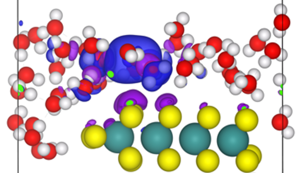
Amylases have been extensively studied in humans, with their 3-dimensional structures characterised in many situations alongside substrates, ligands and inhibitors.13-14 However, structural characteristics of amylases produced by the honeybee (Apis mellifera) are those predicted by the artificial intelligence modelling system, Alphafold,5 based on the existing structures of other amylases.11 We know that calcium and chloride play a significant role in the overall structure of the enzyme.15 From human models, it has been determined that amylases can be classified as metalloenzymes with calcium being key to their stability. They bind to a combination of carbonyl oxygens, an acid residue and water molecules in an 8-coordinate geometry.16 The allosteric binding of chlorine has been shown to encourage calcium binding. Additionally, it induces changes to the conformation of the active site, allowing it to accept substrate. In particular, chlorine attaches to three basic amino acids, which become positively charged under the correct conditions. This induces glutamate to move into the active site so it can participate in the hydrolysis of starch. For these reasons, the activity of amylase is heavily dependent on chlorine concentration.17
Invertase
One of the primary components of nectar is sucrose. This is split into individual glucose and fructose units by invertase. The structure of honeybee invertase has not been determined but a plant invertase, determined by X-ray crystallography,18 is given in Fig. 4. The suggested reaction mechanism for the hydrolysis of sucrose by invertase is shown in Fig. 5.18-19


Like amylases, amino acids with acidic side chains facilitate this reaction as nucleophiles and proton donors. The conjugate base of an acidic amino acid is presumed to act as a nucleophile and attacks the anomeric carbon of the fructose subunit. The nucleophilic attack pushes electrons to the oxygen in the glycosidic bond.
The oxygen attacks the hydrogen on one of the acidic residues, creating a molecule of glucose and a transition state of fructose bound to the enzyme. Water is deprotonated by the resultant negatively charged acid group. The newly created nucleophile attacks the anomeric carbon of the bound fructose, breaking the bond between the enzyme and the fructose.18-19 Related to invertases are sucrases but what distinguishes these is that sucrases break the C1-O bond to glucose, leaving the glucose bound to the enzyme, rather than the O-C2 bond to fructose.20
Proteins are comprised of various substructures, from helical secondary structures to molecular assemblies of quaternary structures. Invertase is an example of an 8-subunit quaternary structure classed as a tetramer of dimers. To produce the oligomer, the enzyme’s monomer undergoes a two-fold rotation to create a dimer and this dimer undergoes a four-fold rotation creating a tetramer of dimers. These protein aggregates are stabilised by hydrogen bonding, disulfide bonds between cysteine residues and interactions between residues of similar hydrophobicity or hydrophilicity.18,21
Glucose oxidase
Many compounds have bactericidal properties. The most well-known of these is hydrogen peroxide, which oxidises the cell wall, destroying the structural integrity of the cell. Additionally, acidity prevents bacterial growth. For these reasons, bees utilise an enzyme that facilitates the production of peroxide and gluconic acid from glucose, available in abundance in honey. This prevents bacterial growth and aids in the maintenance of quality in the honey. 22-23 The predicted structure of glucose oxidase from the honeybee24 is shown in Fig. 6. The predicted mechanism for the oxidation of glucose by glucose oxidase is shown in Fig. 7.22-23,25


In this reaction, the anomeric hydroxyl attacks an oxygen molecule. This creates a trioxide intermediate. The outermost oxygen, carrying a negative charge, bonds to the H of the anomeric hydroxyl restoring electron density to the positively charged anomeric oxygen. Nitrogen, from a histidine residue located in the enzyme’s active site, removes the anomeric hydrogen and thus becomes positively charged. The electrons from the bond that was broken shift to form a double bond to the anomeric oxygen, liberating peroxide ion. The newly formed oxygen nucleophile removes the hydrogen on the positively charged nitrogen in histidine. The result of this reaction is glucono-1,5-lactone and hydrogen peroxide. The lactone product is in equilibrium with gluconic acid. In the dehydrating honey environment, the equilibrium favours the lactone, only forming gluconic acid upon dilution.22-23,25
For every oxidation reaction, there is a converse reduction reaction. The oxidation of glucose is supported by a molecule called flavin adenine dinucleotide (FAD). It features the double purine ring, adenosine attached to a ribose sugar linked to a flavonoid via a diphosphate. FAD is a coenzyme utilised for its reducing potential and its ability to accept electrons in the form of hydride.26 Alongside glucose oxidase, it has been reported that a small amount of glucose dehydrogenase exists in honey. Instead of using oxygen for its oxidoreduction reaction, it makes use of natural electron acceptors such as pyrroloquinoline quinone (PQQ).27
Catalase
Many organisms utilise chemical signalling as an indicator of environmental stress. One such example is hydrogen peroxide. The enzyme catalase is able to break down hydrogen peroxide into water and oxygen, to regulate levels in response to cellular stress.28 As hydrogen peroxide is useful in preventing bacterial growth in honey, catalase is not deliberately added to honey by bees. However, it ends up in the final mixture due to a major component in the honey’s composition - pollen.2-3
Each pollen grain produces catalase to regulate hydrogen peroxide as a means of responding to external pressures in order to protect the cell which is required for reproduction.27 As a result, catalase ends up in honey as an unavoidable side effect of incorporating pollen into the mixture. While levels differ depending on floral origin, catalase activity has not been observed to be high enough to significantly reduce the amounts of hydrogen peroxide produced by glucose oxidase.29 The predicted structure of catalase from honeybee30 is shown in Fig. 8. The proposed reaction mechanism for catalase is shown in Fig. 9.31


At the centre of the active site sits a heme group consisting of iron 4-coordinate to a protoporphyrin ring. The iron is in a 3+ oxidation state. One of the oxygens in hydrogen peroxide attacks the iron forming a peroxyl intermediate with iron now in a 4+ oxidation state. Through the movement of hydrogen, a water leaving group is formed and removed, leaving an oxygen double bonded to the iron. The oxygen attacks one of the hydrogens in another molecule of hydrogen peroxide. This results in a hydroperoxyl and the iron complex in resonance between 4+ and 5+ oxidation states.
The oxygen of the hydroperoxyl is attacked by the iron forming a pseudo-5-membered ring intermediate. This rearranges to give water, which leaves, oxygen, in its typical diatomic form, and the heme group with iron back in the 3+ oxidation state. The overall net product of this reaction is one oxygen and two water molecules for every two molecules of peroxide. It is interesting to note that the quaternary structure of the enzyme is a tetramer with, essentially, four active sites accounting for efficient activity.31-32
Conclusions
The addition of enzymes is essential to making honey nutritional for bees and storable for long periods of time. Each enzyme plays one of a wide variety of biochemical roles from the hydrolysis of oligosaccharides to the production and removal of hydrogen peroxide. They utilise metals, electron carriers and structural features to facilitate important reactions in the production of the bee’s food. Even though we cannot see or taste them, without these biological catalysts, we would not have the sweet and sticky treat we call honey.
References
- Manawa Honey. The Wonder of How Bees Make Honey. 2020. https://www.manawahoney.co.nz/the-wonder-of-how-bees-make-honey/#:~:text=Bees%20make%20honey%20in%20a,honey%20and%20finally%20storing%20it (accessed 03/12/2022).
- Erban, T.; Shcherbachenko, E.; Talacko, P.; Harant, K. J. Nat. Prod. 2019, 82 (5), 1217-1226.
- Rossano, R.; Larocca, M.; Polito, T.; Perna, A.M.; Padula, M.C.; Martelli, G.; Riccio, P. PLoS One 2012, 7 (11), e49164.
- Buttstedt, A.; Moritz, R.F.A.; Erler, S. Biol. Rev. 2014, 89 (2), 255-269.
- Skolnick, J.; Gao, M.; Zhou, H.; Singh, S. J. Chem. Inform. Model. 2021, 61 (10), 4827-4831.
- Phadebas Honey Diastase Test Instructions for Use. 2021. https://www.phadebas.com/wp-content/uploads/PHDT-IFU.pdf (accessed 10/11/2022).
- Sigma-Aldrich Assay Procedure for Invertase. https://www.sigmaaldrich.com/NZ/en/technical-documents/protocol/protein-biology/enzyme-activity-assays/assay-procedure-for-invertase (accessed 01/12/2022).
- Abcam Glucose Oxidase Assay Kit. https://www.abcam.com/glucose-oxidase-assay-kit-fluorometric-ab138884.html#:~:text=The%20glucose%20oxidase%20assay%20protocol,microplate%20reader%20at%20576%20nm (accessed 01/12/2022).
- Hadwan, M. H. BMC Biochem. 2018, 19 (1), 7.
- Tiedge, K.; Lohaus, G. Front. Plant Sci. 2018, 9, 622.
- Alpha Amylase. UniProt. https://www.uniprot.org/uniprotkb/Q9U8X5/entry (accessed 25/01/2023).
- Si Jie, L.; Oslan, S. PeerJ 2021, 9, e11315 (accessed 26/11/2022).
- Ramasubbu, N.; Paloth, V.; Luo, Y.; Brayer, G. D.; Levine, M. J. Acta Cryst. D Biol. Cryst. 1996, 52 (Pt 3), 435-46.
- Berman, H.M.; Westbrook, J.; Feng, Z.; Gilliland, G.; Bhat, T.N.; Weissig, H.; Shindyalov, I.N.; Bourne, P.E. Nucleic Acids Res. 2000, 28 (1), 235-42.
- Bell, A. An investigation of low diastase activity in mānuka honey. MSc thesis, The University of Waikato, 2022.
- Katz, A.K.; Glusker, J.P.; Beebe, S.A.; Bock, C.W. J. Am. Chem. Soc. 1996, 118 (24), 5752-5763.
- Feller, G.; Le Bussy, O.; Houssier, C.; Gerday, C. J. Biol. Chem. 1996, 271 (39), 23836-23841.
- Lammens, W.; Le Roy, K.; Van Laere, A.; Rabijns, A.; Van den Ende, W. J. Mol. Biol. 2008, 377 (2), 378-85.
- Lammens, W.; Roy, K.; Schroeven, L.; Van Laere, A.; Rabijns, A.; Van den Ende, W. J. Exp. Bot. 2009, 60, 727-40.
- Huber, R. E.; Mathison, R. D. Can. J. Biochem. 1976, 54 (2), 153-64.
- Sainz-Polo, M.A.; Ramírez-Escudero, M.; Lafraya, A.; González, B.; Marín-Navarro, J.; Polaina, J.; Sanz-Aparicio, J. J. Biol. Chem. 2013, 288 (14), 9755-9766.
- Bankar, S. B.; Bule, M.V.; Singhal, R.S.; Ananthanarayan, L. Biotech. Adv. 2009, 27(4), 489-501.
- Wohlfahrt, G.; Witt, S.; Hendle, J.; Schomburg, D.; Kalisz, H.M.; Hecht, H.J. Acta Crysta. D Biol. Crysta. 1999, 55 (Pt 5), 969-77.
- Glucose Oxidase. UniProt. https://www.uniprot.org/uniprotkb/Q9U8X6/entry (accessed 25/01/2023).
- Kornecki, J.F.; Carballares, D.; Tardioli, P.W.; Rodrigues, R.C.; Berenguer-Murcia, Á.; Alcántara, A.R.; Fernandez-Lafuente, R. Cat. Sci. Tech. 2020, 10 (17), 5740-5771.
- National Center for Biotechnology Information (2022). PubChem Compound Summary for CID 643975, Flavin adenine dinucleotide. https://pubchem.ncbi.nlm.nih.gov/compound/Flavin-adenine-dinucleotide (accessed 04/12/2022).
- Ferri, S.; Kojima, K.; Sode, K. J. Diabetes Sci. Technol. 2011, 5 (5), 1068-76.
- Yang, T.; Poovaiah, B.W. Proc. Nat. Acad. Sci. 2002, 99 (6), 4097-4102.
- Weston, R. J. Food Chem. 2000, 71 (2), 235-239.
- Catalase. UniProt. https://www.uniprot.org/uniprotkb/A0A7M7L745/entry (accessed 25/01/2023).
- Meunier, B. In Comprehensive Coordination Chemistry II (Eds.: McCleverty, J.A.; Meyer, T.J.), Pergamon: Oxford, 2003, 261-280.
- Hara, I.; Ichise, N.; Kojima, K.; Kondo, H.; Ohgiya, S.; Matsuyama, H.; Yumoto, I. Biochem. 2007, 46 (1), 11-22.